


CORNEAL TOPOGRAPHY
 Corneal
topography is the most accurate tool available for
measuring corneal curvature. It produces a map of the
surface of the cornea and provides essential information
about the cornea's shape, distortions, and astigmatism for
patients considering refractive surgery or who are suffering
from keratoconus.
Corneal
topography is the most accurate tool available for
measuring corneal curvature. It produces a map of the
surface of the cornea and provides essential information
about the cornea's shape, distortions, and astigmatism for
patients considering refractive surgery or who are suffering
from keratoconus.
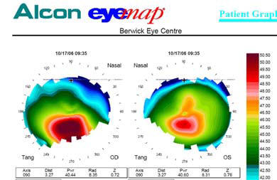
At Berwick Eye Centre we use the Alcon
EyeMap® EH- 290, a state-of-the-art corneal topographer from
one of the leading manufacturers of ophthalmic instruments.
The EyeMap machine may look
intimidating with its black and- white spiral, but don't
worry -- it's a fast, easy and painless test. All you have
to do is place your hands on the table, rest your chin on
the ledge and lean your head on the bar.
The technician will ask you to blink a
few times so the natural tear film on your eye is as uniform
as possible, and then to open your eye wide. The machine
will buzz a little and snap a picture.
You'll see bright
rings of light, and that's all. Those light rings are
shining into your eye and reflecting back into the EyeMap
machine.
Distortions in your cornea bend the
rays of light, so when they bounce back, they arrive in
slightly different positions. Computer software analyses the
shifted light, calculates the surface characteristics of
your cornea, and produces a highly detailed image. Within a
few seconds you'll be able to see the image on the monitor.
Different colours
represent irregularities on the surface of the cornea like
mountains and plains in a landscape. Blue and green indicate
flatter sections of your cornea, while pink and red signify
steeper areas
Corneal
topography is a process for mapping the surface curvature of
the cornea, similar to making a contour map of land.
The cornea is a clear membrane that
covers the front of the eye
and is responsible for about 70 percent of the
eye's focusing power. To a large extent, the shape of the
cornea determines the visual ability of an otherwise healthy
eye. A perfect eye has an evenly rounded cornea, but if the
cornea is too flat, too steep, or unevenly curved, less than
perfect vision results. The purpose of corneal topography is
to produce a detailed description of the shape and power of
the cornea. Using computerized imaging technology, the
3-dimensional map produced by the corneal topographer aids
an ophthalmologist in the diagnosis, monitoring, and
treatment of various visual conditions.
How does corneal
topography work?
The corneal topographer is made up of a
computer linked to a lighted bowl that contains a pattern of
concentric rings. The patient is seated in front of the bowl
with his or her head pressed against a bar while a series of
data points are generated on a Placido Disk, which has been
projected on the cornea. Computer software digitizes these
data points to produce a printout of the corneal shape,
using different colors to identify different elevations. The
procedure itself is painless and brief. It is a noncontact
examination that photographs the surface of the eye using
ordinary light.
The greatest advantage of corneal
topography is its ability to detect conditions invisible to
most conventional testing.
What are the uses of corneal topography?
Corneal topography is not a routine
test. Rather, it is used in diagnosing certain types of
problems, in evaluating a disease's progression, in fitting
some types of contact lenses, and in planning surgery. It is
commonly used in preparing for refractive eye surgery. The
corneal topography map is used in conjunction with other
tests to determine exactly how much corneal tissue will be
removed to correct the visual defect.
Corneal topography is used in the diagnosis and management
of
various corneal curvature abnormalities and diseases
such as:
•
Diagnosis of hidden astigmatism
•
Keratoconus, a degenerative condition that
causes a thinning of the cornea
•
Corneal transplants
•
Corneal scars or opacities
•
Corneal deformities
•
Fitting contact lenses
•
Irregular astigmatism following corneal
transplantation
•
Planning cataract surgery
•
Planning refractive surgery